Dr. Ashish Mitra (Cataract & Retina Surgeon)
Ratan Jyot Netralaya, 49/19B Near Manmohan Park Chauraha, Katra, Prayagraj - 211001
CONTACT
+91 8085309909
+91 8085309909
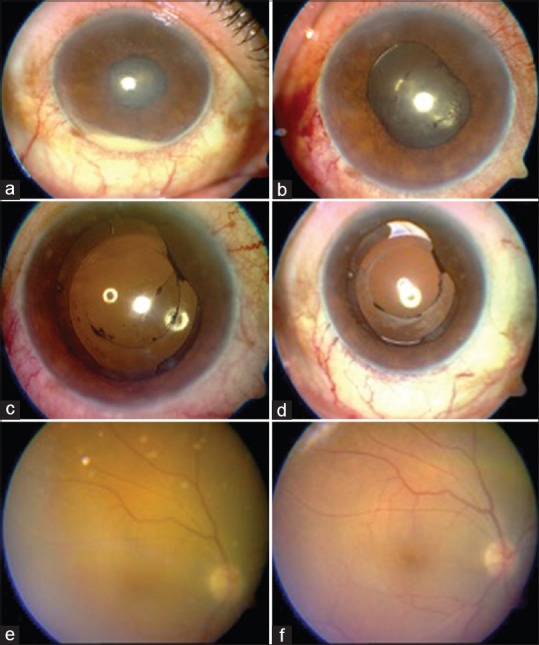
Endophthalmitis :
Endophthalmitis can be divided into two main types based on the mode of infection as: Exogenous and Endogenous (ie, metastatic). Exogenous endophthalmitis results from direct inoculation as a complication of ocular surgery, foreign bodies, blunt or penetrating ocular trauma. Endogenous endophthalmitis results from the hematogenous spread of organisms from a distant source of infection (eg, endocarditis). Endogenous endophthalmitis is quite rare. Various studies have reported incidence varying from 2% to 15%. In endogenous endophthalmitis, blood-borne organisms permeate the blood-ocular barrier either by direct invasion (septic emboli) or by changes in vascular endothelium caused by substrates released during infection. Destruction of intraocular tissues may be due to direct invasion by the organism and/or from inflammatory mediators of the immune response. Due to increase in the spread of AIDS, more frequent use of immunosuppressive agents, and the use of more invasive procedures, the patients at risk of endogenous endophthalmitis are increasing .
Endophthalmitis as a complication of vitreoretinal surgery is relatively uncommon. Scleral buckle infection usually appears between the second and seventh day of surgery, and, presents as severe lid oedema with conjunctival chemosis associated with severe ocular pain and headache. Intraocular inflammation may develop associated with media haziness, subretinal exudation and localised exudative retinal detachment. In delayed subacute cases fistula and granuloma form, resulting in exposure of the buckle. Diabetes and prolonged duration of surgery have been identified as specific risk factors in this setting. Incidence of endophthalmitis following pars plana vitrectomy has been reported as low as 0.051%.
HOW TO EXAMINE A CASE OF ENDOPHTHALMITIS ?
The examination of the suspected patient should include a detailed history, visual acuity assessment, examination of ocular adnexa (any sign of lid swelling), anterior chamber evaluation (using a slit lamp) and examination of the vitreous.
Associated signs like leaking filtering blebs, wound leak etc. should be noted if any. Fundus view may be obscured because of anterior chamber reaction or due to vitreous exudates. The clarity of ocular media could be assessed by indirect ophthalmoscopy.
For more information or to book appointment for Endophthalmitis Treatment in Prayagraj, Please Contact Dr. Ashish Mitra at +91 8085309909 or write us at info@eyeclinicprayagraj.com